Horses Inside Out Conference 2020 - Anatomy In Action - Part 7
Hello and welcome back to my blog series on the Horses Inside Out Conference - Anatomy In Action.
Today I am very excited to share with you the research vet Sue Dyson presented to us on day one of the conference (we were lucky enough to have presentations from Sue on both days).
Sue is a world renowned expert in equine orthopaedics, with a particular interest in poor performance and subtle and complex lameness in sport horses.
Sue an expert in diagnostic imaging, radiography, ultrasonography, scintigraphy and magnetic resonance imaging.
Sue lectures internationally and has published many books and more than papers in scientific journals.
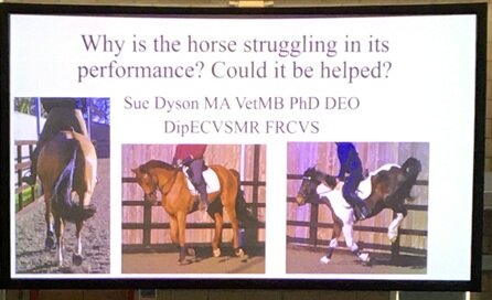
This presentation was discussing the reasons why a horse may be struggling in performance and could it be helped?
Sue began by discussing with us how many training and performance problems have historically been attributed to the horse’s behaviour or the rider’s skill.
However there is increasing recognition that musculoskeletal pain may be the underlying problem either primary or secondary to:
An ill fitting saddle for the horse
An ill fitting saddle for the rider
Inappropriate training methods
Failure to develop key muscle groups
Some of my own personal key notes from the presentation are below, I hope these are useful to you:
With head and neck up it is physically impossible for the horse to move through the back
Never underestimate how saddle fit can affect head and neck carriage, stride length and movement through the Thoracolumbosacral region (the part of the spine we call the back, lower back and pelvis)
The width of the horse’s back increases during exercise - we must fit saddles with this in mind
A dry patch in the horse’s coat after exercise means the saddle is too tight
The horse’s hind limbs cannot come under them if there is lack of flexion in the sacroilliac joint (pelvis)
Sue then shared with us the development and validation of the Ridden Horse Ethogram. This is a powerful tool for the recognition of musculoskeletal pain.
Essentially (please note these are my own words and my own understanding of the presentation) The Ridden Horse Ethogram is a list of 24 behaviours displayed by a horse that may indicate pain.
These include:
ears back
mouth opening
tongue out
change in eye posture and expression
going above the bit
head tossing
tilting the head
unwillingness to go
crookedness
hurrying
changing gait spontaneously
poor quality canter
resisting
stumbling
toe dragging
A horse that displays more than 8 of these 24 behaviours may have low grade lameness that we may otherwise miss without using this diagnostic tool.
The Ridden Horse Ethogram may be a useful tool enabling earlier recognition of lameness and avoidance of punishment-based training.
Such fascinating work, and a useful tool we can all use to evaluate those ‘tricky’ horses that may be behaving as such as they have an underlying lameness we have been unable to spot just by watching them move.
I would like to leave you with a final thought, something that came up during the time at the end of the presentation when delegates had the opportunity to ask Sue some questions.
Sue described how bone scanning can show many false positives and negatives when used as a diagnostic tool for lameness.
She believes nerve blocks followed by radiography is a much better course of action and bone scanning can at times be a waste of client’s money.
Certainly food for thought!
Thank you so much for visiting my blog, I hope you have found it useful.
There are lots of resources online where you can find out more about Sue and her research.
Thank you again to Horses Inside Out for allowing me access to their professional images. Although many of the images in this blog are my own screen shots of Sue’s fabulous slides.
In my next blog I will be sharing with you the presentation by farrier Mark Johnson.
Thanks so much


